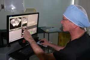
Contributed by Dr. Kia Michel –
Diagnosing prostate cancer starts with regular prostate screening and talking to your doctor about symptoms and family history. This much we know.
So what happens after you are screened for prostate cancer and you know what your PSA (prostate specific antigen) is?
Mostly likely if you have an elevated PSA or you have had your PSA tested over time and there has been a big jump (like from 2 to 4 in just one year) your physician will recommend that you have a biopsy. A prostate biopsy is a procedure in which small samples of the prostate are removed and then looked at under a microscope by a pathologist.
The traditional type of biopsy used to diagnose prostate cancer is a core needle biopsy. It is usually done by an urologist as a 10 -20 minute procedure in a doctor’s office. The urologist uses a transrectal ultrasound probe to help guide the insertion of thin hollow needles that are placed through the wall of the rectum into the prostate. When the needle is pulled out, it removes a small amount of prostatic tissue. This is typically repeated about 12 times targeting different areas of the prostate.
However, some urologists believe there is a better; more accurate and effective way to offer patients biopsies. The core needle biopsy that has been in practice since the 1980s doesn’t really allow doctors to determine where the tumors might be in the prostate so they are really just randomly deciding which tissue to exact.
Another option is something called a fusion biopsy where a urologist uses MRI images to better locate the areas in the prostate where the tumor may be to guide the biopsy. An MRI scan is better at revealing changes in tissue in the prostate gland. Although a doctor cannot diagnose prostate cancer from the MRI image, it can be used to identify suspicious areas that warrant closer examination with a needle biopsy.
Advances in technology now allow these MRI images to be combined with live, real-time ultrasound images of the prostate. A patient first undergoes the MRI scan. A radiologist reviews it and marks suspicious areas. Later, when the patient returns for the actual biopsy, an ultrasound probe is placed in the rectum and as the probe moves around, the fusion software overlays the MRI images on the live ultrasound images giving 3D ultrasound/MRI view of the prostate gland. This Fused image is then used to guide the needle biopsy and now the physician has a clear roadmap of where to target versus just randomly inserting needles.
By being able to more accurately diagnose prostate cancer with a fusion biopsy, urologists also have more options for non-invasive treatments that reduce treatment time, recovery time and overall risk of side effects such as impotence and incontinence.
HIFU, also know as high intensity focused ultrasound, for prostate cancer is an incredibly targeted therapy that enable the physician to target specifically where the cancer is and not cause collateral damage to the area. This means that is a doctor can use MRI fusion biopsy to diagnose the exact location of the cancer; they can use HIFU to treat it with few side effects and a quick recovery.
The patient experience below may help to fully understand how MRI fusion and HIFU complement each other and allow physicians to offer men a minimally invasive prostate cancer treatment.
Treatment Review
A patient had a PSA of 3.9 and was then scheduled for a MRI followed by a fusion biopsy. The MRI showed suspicious areas on both sides of the gland.

The biopsy was completed using the UroNav fusion biopsy system, and the results confirmed that there was cancer on both the left and right side of the glade.
After discussing with the patient it was determined that HIFU was a good option because of its ability to achieve cancer control without a huge risk of complications.
A post treatment MRI was planned as well so the treating physician could reevaluate knowing HIFU doesn’t prevent a patient from having more treatment if needed.

The decision was made to do a urethral sparing HIFU treatment in order to reduce risk of retention and incontinence.
A urethral sparing HIFU treatment means that the physician destroys tissue on either side of the urethra (which runs directly through the prostate) but does not ablate or destroy the prostatic urethra.
The red in the image indicates areas where HIFU is delivered and tissue is destroyed.
A few weeks after the treatment, another MRI was done that confirmed that all the targeted tissue and cancer was destroyed.

Conclusion
MRI fusion prostate biopsies are becoming increasingly popular among men who are worried about over treating a prostate cancer that may be slow growing. It helps doctors understand exactly what they are looking for and serves as a type of GPS for finding the cancer. Once they know where the cancer is they can offer treatment options that are customized for their patient.
About the Author
 Dr. Kia Michel of Prostate Cancer Specialists of Los Angeles specializes in offering patients MRI-Guided biopsies as well as focal HIFU therapy. He is a leading prostate cancer expert in Los Angeles as well as an acclaimed cancer and robotic surgeon that has been one of the pioneers of prostate focal therapy in Southern California. Dr. Michel has expertise in MRI fusion prostate biopsies and prostate mapping. This acumen has allowed him to champion focal therapy with HIFU for patients with localized prostate cancer. His expertise is sought after by patients nationally and internationally. Dr. Michel is highly respected by his peers and serves as an expert consultant to his colleagues. Read more about Dr. Michel or schedule a consultation with him here.


Comments are closed.